Muscles of the Anterior Abdominal Wall
Introduction
Six pairs of abdominal muscles
From superficial to deep they are :-
Rectus abdominis muscle
External oblique muscle
Internal oblique muscle
Transversus abdominis muscle
Quadratus lumborum muscle (posterior abdominal muscle)
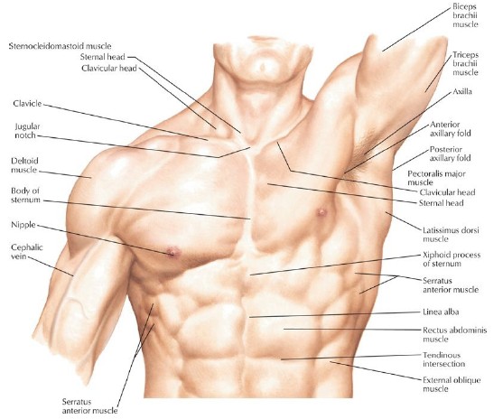
Linea alba
Strong midline tendinous cord - extends from xiphisternum to the symphysis pubis
Rectus abdominis
Rectus means straight
One on each side of the linea alba
Direction of the fibres : vertical
Most superficial muscle
Broad and flat
Origin : transvers part of the pubic bone Insertion : lower ribs and xiphisternum - attached to linea alba
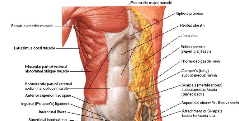
External oblique
Extends from the lower ribs to the iliac crest and attaches medially to the linea alba
Direction of the fibres : downwards and forwards
Laterally muscular and medially aponeurotic
Internal oblique
Deep to the external oblique
Arises from the iliac crest and lumbar spines
Direction of the fibres : upwards towards the midline, at right angles to the fibres of the external oblique
Attached to the lower ribs and linea alba
Transversus abdominis
The deepest layer of muscles
Arises from the iliac crest and the lumbar vertebrae
Fibres pass across the abd. wall to linea alba
Direction of the fibres : transverse; at right angle to the rectus abdominis
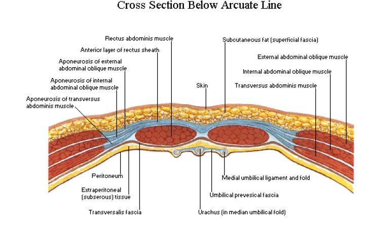
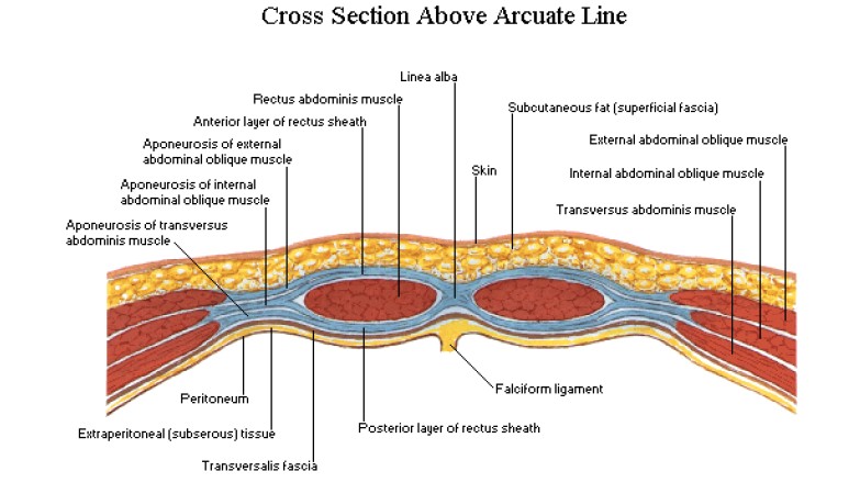
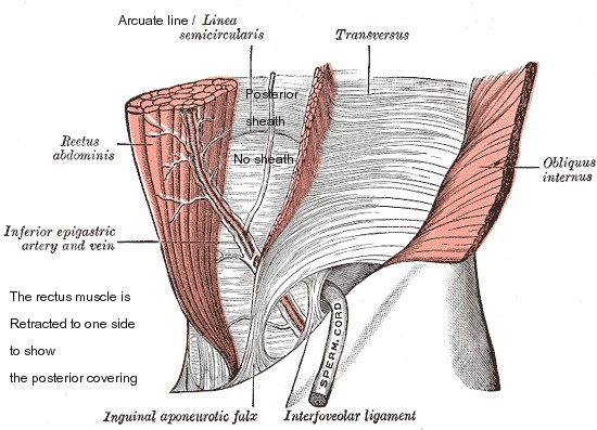
Rectus Sheath
The aponeurotic sheaths of the External oblique muscle, the Internal oblique muscle and the Transversus abdominis muscle fuse medially near the lateral border of the Rectus abdominis muscle and then split into two to enclose the Rectus abdominal muscle and then fuse again at the midline to form linea alba.
The splitting of the fused aponeuroses of these three muscles occurs from the xiphoid process to upto a point below the umbilicus;
After that point there is no splitting; all the three aponeuroses pass anterior to the rectus abdominis muscle.
So at this point onwards the recus abdominis muscle is not covered posteriorly by any fibrous sheath.
There is horizontally crescentic line marking the end of the posterior covering/posterior rectus sheath - Called Arcuate Line
Functions
Forms a strong wall for the abdominal contents
Compressing the abdominal organs
Flex the vertebral column in the lumbar region
Rotation of lower vertebrae
Lateral flexion of the trunk